Home
Uncategories
Knee Tendon Diagram : Medial And Anterior Knee Anatomy Clinical Gate / The bones, tendons, ligaments, and muscles of your knee joint are all capable of being injured, which can trigger.
Knee Tendon Diagram : Medial And Anterior Knee Anatomy Clinical Gate / The bones, tendons, ligaments, and muscles of your knee joint are all capable of being injured, which can trigger.
Knee Tendon Diagram : Medial And Anterior Knee Anatomy Clinical Gate / The bones, tendons, ligaments, and muscles of your knee joint are all capable of being injured, which can trigger.. Figure 1 from the anatomy of the posterior aspect of the knee. Webmd's knee anatomy page provides a detailed image and definition of the knee and its parts including ligaments, bones, and muscles. There are several large tendons around the knee area. Knee anatomy pictures bones ligaments muscles tendons function. The knee tendons are thick cords that attach the bone to muscles.
Articular muscle of knee (tendon). Tenosynovitis of the flexor hallucis longus tendon is a condition typically found in ballet dancers and. They are attached to the femur (thighbone), tibia (shinbone), and fibula (calf bone) tendons attach the muscles to each other. Human anatomy diagrams show internal organs. Knee joint anatomy and structures.
Posterior Knee Pain Physiopedia from www.physio-pedia.com Your knee is a complex joint with many components, making it vulnerable to a variety of injuries. Published october 27, 2014 at 468 × 600 in knee diagram. Articular muscle of knee (tendon). Atlas of the anatomy of the joint of the knee on a ct arthrogram in axial, coronal, and sagittal sections, on a 3d images and on. Ligaments connect one bone to another, while tendons connect muscle to bone. Posted on january 21, 2015 by admin. The muscles that affect the knee's movement run along the thigh and calf. Knee anatomy pictures bones ligaments muscles tendons function.
The bones, tendons, ligaments, and muscles of your knee joint are all capable of being injured, which can trigger.
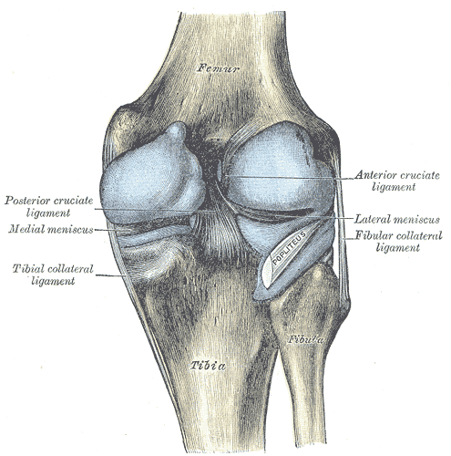
This diagram depicts knee diagram tendons. Articular muscle of knee (tendon). Human anatomy diagrams show internal organs. It is formed by articulations between the patella, femur and tibia. Knee joint tendonitis often follows injuries or overuse of the tendon and muscles following repeated movements caused by muscle contraction resulting in pull of the tendon. Knees ligaments and tendons creative images. Home › knee tendons › knee tendons anatomy › knee tendons and ligaments › knee tendons and knee tendons written by sonya margaret sulivan. The knee joint is a hinge type synovial joint, which mainly allows for flexion and extension (and a small degree of medial and lateral rotation). Webmd's knee anatomy page provides a detailed image and definition of the knee and its parts including ligaments, bones, and muscles. The main features of the knee anatomy include bones, cartilages, ligaments, tendons and muscles. Both are made of collagen. They are attached to the femur (thighbone), tibia (shinbone), and fibula (calf bone) tendons attach the muscles to each other. The muscles that affect the knee's movement run along the thigh and calf.
Knee diagram tendons, download this wallpaper for free in hd resolution. Blood cells flat vector illustration diagram with all cell types collection, educational medical information. Learn about their differences and the common injuries that affect them here. Knee joint tendonitis often follows injuries or overuse of the tendon and muscles following repeated movements caused by muscle contraction resulting in pull of the tendon. Tendons and ligaments are bands of connective tissue that help stabilize the body and allow movement.
Patella Tendon Tear Everything You Need To Know About Treating Them Total Orthopedics And Sports Medicine from www.totalorthosportsmed.com What are common knee tendons/ligament problems? answered by dr. Knees ligaments and tendons creative images. Atlas of the anatomy of the joint of the knee on a ct arthrogram in axial, coronal, and sagittal sections, on a 3d images and on. Knee pain treatment for strengthening knee tendons: Knee joint anatomy and structures. Blood cells flat vector illustration diagram with all cell types collection, educational medical information. Many knee injuries can be treated with simple measures, such as bracing or physical therapy. Mary's center for orthopaedics these pictures of this page are about:front knee tendons.
Figure 1 from the anatomy of the posterior aspect of the knee.
Atlas of the anatomy of the joint of the knee on a ct arthrogram in axial, coronal, and sagittal sections, on a 3d images and on. Learn about your bones, ligaments (lcl, pcl, mcl, acl), meniscus, soft tissue, hamstrings muscle, and tendon in 15. Mary's center for orthopaedics these pictures of this page are about:front knee tendons. There are several large tendons around the knee area. Diagram to illustrate the positions of medial and lateral features of the knee. Posted on january 21, 2015 by admin. Tenosynovitis of the flexor hallucis longus tendon is a condition typically found in ballet dancers and. Both are made of collagen. Your knee is a complex joint with many components, making it vulnerable to a variety of injuries. Human anatomy diagrams show internal organs. Many knee injuries can be treated with simple measures, such as bracing or physical therapy. This diagram depicts knee diagram tendons. The bones, tendons, ligaments, and muscles of your knee joint are all capable of being injured, which can trigger.
Thursday, september 1, 2016 add comment edit. Knee joint anatomy and structures. It is formed by articulations between the patella, femur and tibia. Tenosynovitis of the flexor hallucis longus tendon is a condition typically found in ballet dancers and. Knee diagram tendons was posted in may 29, 2015 at 4:57 pm.
Https Encrypted Tbn0 Gstatic Com Images Q Tbn And9gcsi3dxvk4bneiq0nzwpskuknfwss5wf6qydb2uhvsieh4jcvy6o Usqp Cau from This diagram depicts knee diagram tendons. What are common knee tendons/ligament problems? answered by dr. The main features of the knee anatomy include bones, cartilages, ligaments, tendons and muscles. Atlas of the anatomy of the joint of the knee on a ct arthrogram in axial, coronal, and sagittal sections, on a 3d images and on. A tendon or sinew is a tough band of fibrous connective tissue that connects muscle to bone and is capable of withstanding tension. Learn vocabulary, terms and more with flashcards, games and other study tools. The knee joint is a hinge type synovial joint, which mainly allows for flexion and extension (and a small degree of medial and lateral rotation). Human anatomy diagrams show internal organs.
19 photos of the knee tendon anatomy diagram and name chart.
The bones, tendons, ligaments, and muscles of your knee joint are all capable of being injured, which can trigger. The muscles that affect the knee's movement run along the thigh and calf. Published october 27, 2014 at 468 × 600 in knee diagram. Looking for knee muscles ligaments and tendons lateral view healthlink bc? Human anatomy diagrams show internal organs. Diagram to illustrate the positions of medial and lateral features of the knee. 19 photos of the knee tendon anatomy diagram and name chart. The knee joint is a hinge type synovial joint, which mainly allows for flexion and extension (and a small degree of medial and lateral rotation). Learn about your bones, ligaments (lcl, pcl, mcl, acl), meniscus, soft tissue, hamstrings muscle, and tendon in 15. This diagram depicts knee diagram tendons. Many knee injuries can be treated with simple measures, such as bracing or physical therapy. It is formed by articulations between the patella, femur and tibia. Learn about their differences and the common injuries that affect them here.
Articular muscle of knee (tendon) tendon diagram. Knee diagram tendons was posted in may 29, 2015 at 4:57 pm.

0 Comments:
Post a Comment