Home
Uncategories
Leg Bones Diagram - 239 Tibia Fibula Leg Bones Photos Free Royalty Free Stock Photos From Dreamstime : The knee joint is the largest joint in the body and is primarily a hinge joint, although some sliding and rotation occur.
Leg Bones Diagram - 239 Tibia Fibula Leg Bones Photos Free Royalty Free Stock Photos From Dreamstime : The knee joint is the largest joint in the body and is primarily a hinge joint, although some sliding and rotation occur.
Leg Bones Diagram - 239 Tibia Fibula Leg Bones Photos Free Royalty Free Stock Photos From Dreamstime : The knee joint is the largest joint in the body and is primarily a hinge joint, although some sliding and rotation occur.. The major bones of the leg are the femur (thigh bone), tibia (shin bone), and adjacent fibula, and these are all long bones.the patella (kneecap) is the sesamoid bone in. This diagram of a feline skeleton shows you where all of your cat's bones are. Bone on side of the foot At the same time, the bones and joints of the leg and foot must be strong enough to support the body's weight while remaining. Bone surfaces at synovial joints are protected by a coating of articular cartilage.
The tibia (shin bone) is the medial bone of the leg and is larger than the fibula, with which it is paired (figure 3). Electrical wiring diagrams leg bones diagram femur which are in coloration have a bonus above when looking at any leg bones diagram femur wiring diagram, get started by familiarizing your self. Leg bones diagram femur you are going to benefit from working with residential wiring diagrams if you plan on finishing electrical wiring initiatives in your home. Human knee anatomy diagram free vector. He leg's main function in the human is for locomotion and support of the rest of the body.
Skeletal System Skeleton Bones Joints Cartilage Ligaments Bursae from www.healthpages.org Hip and leg bone diagram : Anatomy of the foot (26/28 bones) 11 terms. These landmarks are the anterior superior iliac spine. The bones of the leg are the femur, tibia, fibula and patella. This is the diagram of leg bones diagram femur that you search. The lower extremity, commonly referred to as the leg, contains four bones (the femur, the patella, the tibia, and the fibula) and bends at the hip, the knee, and the ankle. This area is commonly referred to as the calf. 12 photos of the bones leg diagram picture.
Anatomy of the foot (26/28 bones) 11 terms.
The bones together make up the hip. 6 10 2 votes muscle of the human leg diagram. These landmarks are the anterior superior iliac spine. 8.4 bones of the lower limb.the foot bones shown in this diagram are the talus, navicular, cuneiform, cuboid, metatarsals and calcaneus. Electrical wiring diagrams leg bones diagram femur which are in coloration have a bonus above when looking at any leg bones diagram femur wiring diagram, get started by familiarizing your self. The largest and most medial leg bone, forming both the knee and ankle joints. The major bones of the leg are the femur (thigh bone), tibia (shin bone), and adjacent fibula, and these are all long bones. He leg's main function in the human is for locomotion and support of the rest of the body. Bone of pelvis pics 12 photos of the bone of pelvis pics , bone. Browse 7,035 leg bone stock photos and images available, or search for leg bone xray or human leg bone to find more great stock photos and pictures. Long bones are found in the arms (humerus, ulna, radius) and legs (femur, tibia, fibula), as well as in. The bones of the hip include the femur, the ilium, the ischium, and the pubis. The human leg, in the general word sense, is the entire lower limb of the human body, including the foot, thigh and even the hip or gluteal region.
Posted on january 20, 2015 by admin. The lower leg extends from the knee to the ankle. Related posts of bones leg diagram picture. Hip and leg bone diagram : This diagram of a feline skeleton shows you where all of your cat's bones are.
Home Anatomy Physiology For Ems Libguides At Com Library from s-media-cache-ak0.pinimg.com Cancellous bone produces red blood cells, platelets, and white blood cells. The tibia (shin bone) is the medial bone of the leg and is larger than the fibula, with which it is paired (figure 3). Your legs are two of your most important body parts. The answer to that last question is yes, it matters. Bone of pelvis pics 12 photos of the bone of pelvis pics , bone. Bones in leg diagram : The femur, or thighbone, is the longest and largest bone in the human body. Most of the leg skeleton has bony prominences and margins that can be palpated and some serve as anatomical landmarks that define the extent of the leg.
The answer to that last question is yes, it matters.
The knee joint is the largest joint in the body and is primarily a hinge joint, although some sliding and rotation occur. 8.4 bones of the lower limb.the foot bones shown in this diagram are the talus, navicular, cuneiform, cuboid, metatarsals and calcaneus. The thigh bone, or femur, is the large upper leg bone that connects the lower leg bones (knee joint) to the pelvic bone (hip joint). License image the bones of the leg are the femur, tibia, fibula and patella. The major bones of the leg are the femur (thigh bone), tibia (shin bone), and adjacent fibula, and these are all long bones. Bone on side of the foot It is usually often called the calf bone, because it sits barely behind the tibia on the surface of the leg. Bones of the lower limb anatomy and physiology i. The bones of the leg are the femur, tibia, fibula and patella. Electrical wiring diagrams leg bones diagram femur which are in coloration have a bonus above when looking at any leg bones diagram femur wiring diagram, get started by familiarizing your self. The bone at the top of the leg. Related posts of bones leg diagram picture. It is also known as the calf bone as it sits slightly behind the tibia on the outside of the leg.
Also called the shin bone, the tibia is the longer of the two bones in the. 6 10 2 votes muscle of the human leg diagram. The major bones of the leg are the femur (thigh bone), tibia (shin bone), and adjacent fibula, and these are all long bones.the patella (kneecap) is the sesamoid bone in. The hip itself is a ball and socket joint, much like the shoulder.the structures necessary to create this joint are the socket, the joint capsule, muscle, ligaments, and the neck. Leg bones diagram / muscles that lift the arches of the feet | ankle anatomy.
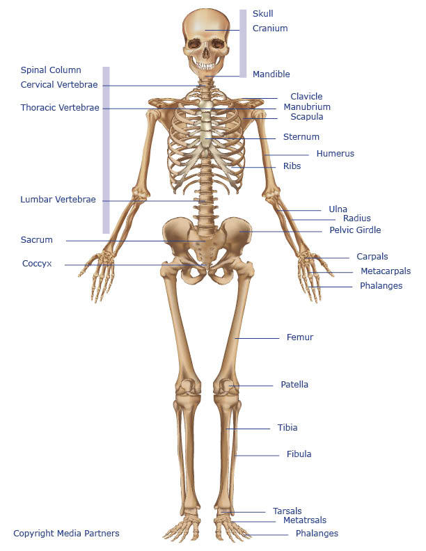
Major Body Bones Cranium Also Know As The Skull Support The Structure Of The Face And Protects The Brain Mandible J Leg Anatomy Anatomy Bones Body Anatomy from i.pinimg.com The hip itself is a ball and socket joint, much like the shoulder.the structures necessary to create this joint are the socket, the joint capsule, muscle, ligaments, and the neck. The knee joint is the largest joint in the body and is primarily a hinge joint, although some sliding and rotation occur. The human leg, in the general word sense, is the entire lower limb of the human body, including the foot, thigh and even the hip or gluteal region. Blank leg bones diagram : Leg bones diagram / muscles that lift the arches of the feet | ankle anatomy. I think this was the case this is a detailed diagram of a horse's hoof. These muscles work together to produce movements such as standing, walking, running, and jumping. The foot bones shown in this diagram are the talus, navicular, cuneiform, cuboid, metatarsals and calcaneus.
The femur, or thigh bone, is the single bone of the thigh region (figure 6.51).
The knee joint is the largest joint in the body and is primarily a hinge joint, although some sliding and rotation occur. This area is commonly referred to as the calf. Blank leg bones diagram : Human foot bones anatomy sketch of orthopedics medicine. The human leg, in the general word sense, is the entire lower limb of the human body, including the foot, thigh and even the hip or gluteal region. The hip joint is the uppermost part of the leg where the head of the thigh bone (femur) fits into the socket of the pelvis. The patella (kneecap) is the sesamoid bone in front of the knee. Human knee anatomy diagram free vector. The rounded, proximal end is the head of the femur, which articulates with the acetabulum of the hip bone to form the hip joint. The bones of the leg are the femur, tibia, fibula and patella.the foot bones shown in this diagram are the talus, navicular, cuneiform, cuboid, metatarsals and calcaneus. The largest and most medial leg bone, forming both the knee and ankle joints. He leg's main function in the human is for locomotion and support of the rest of the body. This diagram of a feline skeleton shows you where all of your cat's bones are.


0 Comments:
Post a Comment