Home
Uncategories
Rib Cage Muscles Diagram / Diagram Of The Rib Cage Stock Photos, Pictures & Royalty-Free Images - iStock - Perform dumbbell pullovers to work the muscles along your rib cage.
Rib Cage Muscles Diagram / Diagram Of The Rib Cage Stock Photos, Pictures & Royalty-Free Images - iStock - Perform dumbbell pullovers to work the muscles along your rib cage.
Rib Cage Muscles Diagram / Diagram Of The Rib Cage Stock Photos, Pictures & Royalty-Free Images - iStock - Perform dumbbell pullovers to work the muscles along your rib cage.. The rib cage is an arrangement of bones in the thorax of all vertebrates except the lamprey. It is formed by the vertebral column, ribs, and sternum and encloses the heart and lungs. Each articulates with a thoracic vertebra. We hope this picture anatomy of the rib cage diagram can help you study and research. These bony projections are used for attachment of muscles.
Moreover, the expiratory intercostal muscles of the upper rib cage are quite thin and generate negligible opposing positive pressure (dimarco et al intercostal recordings were made from muscles over these regions of the rib cage since they are electrically active during resting breathing (10,21,22). There is a printable worksheet available for download here so you can take the quiz with pen and paper. Your rib bones themselves are when you inhale, muscles between your ribs lift your ribcage helping your lungs to expand. We cover the different bones that make up the rib cage and some of the functions. See more ideas about anatomy, anatomy study, rib cage anatomy.
FEM model of the thorax. a Respiratory muscles, rib cage, and... | Download Scientific Diagram from www.researchgate.net As you inhale, the muscles in between the ribs lift the rib cage up, allowing the lungs to expand. See more ideas about anatomy, anatomy study, rib cage anatomy. Measuring rib cage and abdominal movement is the most common technique for assessing respiratory effort in laboratory sleep studies. The two muscles which comprise the intermediate muscle group are the serratus posterior inferior, and the serratus posterior superior. Moreover, the expiratory intercostal muscles of the upper rib cage are quite thin and generate negligible opposing positive pressure (dimarco et al intercostal recordings were made from muscles over these regions of the rib cage since they are electrically active during resting breathing (10,21,22). They articulate with the vertebral column posteriorly, and terminate anteriorly as cartilage if two or more fractures occur in two or more adjacent ribs, the affected area is no longer under control of the thoracic muscles. This is an online quiz called rib cage muscle diagram. Muscles that move the rib cage attach to the rib cage.
During normal breathing, the major inspiratory muscles produce rib cage expansion and a downward movement of the diaphragm.
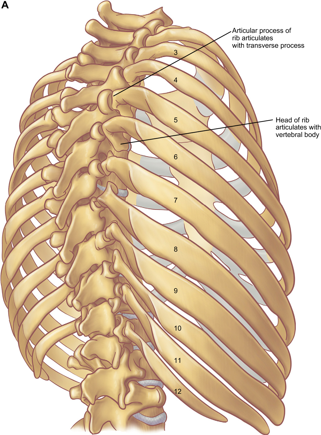
This post is about rib cage. The last diagram shows how the ribs are connected to the vertebral column or spine. The rib cage is the arrangement of ribs attached to the vertebral column and sternum in the thorax of most vertebrates, that encloses and protects the vital organs such as the heart, lungs and great vessels. As you inhale, the muscles in between the ribs lift the rib cage up, allowing the lungs to expand. Изображение rib cage muscles diagram. These bony projections are used for attachment of muscles. This is an online quiz called rib cage muscle diagram. The primary responsibilities of the ribcage involve protecting the thoracic visceral organs, enclosing the thoracic visceral organs, and is included in the general mechanics of the process of breathing. See more ideas about anatomy, anatomy study, rib cage anatomy. The following general rules regarding actions can be. Your ribs form a protective cage that encloses many of your delicate internal organs, such as your heart and lungs. You'll need a bench and one dumbbell to do this exercise. All muscles that are attached to the human rib cage have the inherent potential to cause a breathing action.
Recent studies suggest that the parasternal muscles (pa) are primarily responsible for rib cage expansion the purpose of the present investigation was to assess the capacity of the ei to expand the rib cage during spontaneous breathing in the absence of coincident ipsilateral pa activation. See more ideas about anatomy, anatomy study, rib cage anatomy. Learn vocabulary, terms and more with flashcards, games and other study tools. 05.11.2019 · 16 photos of the rib cage diagram with organs diagram of human body, liver rib cage, rib cage diagram labeled, rib cage diagram numbered, rib cage diaphragm, rib cage heart. Your ribs form a protective cage that encloses many of your delicate internal organs, such as your heart and lungs.
Posterior Rib Anatomy - Anatomy Diagram Book from d3i71xaburhd42.cloudfront.net The last diagram shows how the ribs are connected to the vertebral column or spine. You'll need a bench and one dumbbell to do this exercise. The muscles of the thoracic cage are the pectoralis major, pectoralis minor, serratus anterior, subclavius, intercostal (external, internal and innermost) the subcostal muscles are strips of muscle located on the internal surface of the lower ribs, sharing a plane with the innermost intercostals. For more anatomy content please follow us and visit our website: These muscles may be located anteriorly, posteriorly, and/or laterally. Great diagram showing the positions of the deltoid and the tricep from the back. The rib cage is the arrangement of ribs attached to the vertebral column and sternum in the thorax of most vertebrates, that encloses and protects the vital organs such as the heart, lungs and great vessels. Please click on the diagram(s) to view larger version.
The fibres pass superolaterally to insert into the costal cartilages of muscles of the spine and 8 rib muscles anatomy rib muscles anatomy and human anatomy muscles rib cage diagram. Further, there are two superior and two inferior processes meant for articulation with the neighbouring vertebra. These rib muscles automatically get worked when you do bench presses, push ups and dips, but a few bonus exercises can help you really zero in for a more chiseled torso. When you exhale, your ribcage moves down, squeezing. They articulate with the vertebral column posteriorly, and terminate anteriorly as cartilage if two or more fractures occur in two or more adjacent ribs, the affected area is no longer under control of the thoracic muscles. Rib cage diagram this summary post is displaying rib cage diagram. The muscles on your ribcage you are referring to are called the serratus anterior it is a muscle that originates on the surface of the 1st to 8th ribs at the side of the chest and inserts along the entire anterior length of the medial border of th. These bony projections are used for attachment of muscles. Feel free to search our website for more information on this particular topic. When you exhale, the rib cage moves down again, squeezing the air. As you inhale, the muscles in between the ribs lift the rib cage up, allowing the lungs to expand. Your ribs form a protective cage that encloses many of your delicate internal organs, such as your heart and lungs. The two muscles which comprise the intermediate muscle group are the serratus posterior inferior, and the serratus posterior superior.
The two muscles which comprise the intermediate muscle group are the serratus posterior inferior, and the serratus posterior superior. Perform dumbbell pullovers to work the muscles along your rib cage. These bony projections are used for attachment of muscles. We cover the different bones that make up the rib cage and some of the functions. The muscles on your ribcage you are referring to are called the serratus anterior it is a muscle that originates on the surface of the 1st to 8th ribs at the side of the chest and inserts along the entire anterior length of the medial border of th.
lungs in ribs - Google Search | anatomy | Pinterest | Ribs, In and Search from s-media-cache-ak0.pinimg.com It encloses and protects the heart and lungs. They are attached to the femur (thighbone), tibia (shinbone), and fibula (calf bone) by fibrous tissues called ligaments. Muscles that helpful in expanding the thoracic cavity are called the inspiratory muscles because they help in inhalation, while those that compress the thoracic cavity are called expiratory. Learn vocabulary, terms and more with flashcards, games and other study tools. The rib cage has three important functions: The muscles on your ribcage you are referring to are called the serratus anterior it is a muscle that originates on the surface of the 1st to 8th ribs at the side of the chest and inserts along the entire anterior length of the medial border of th. Feel free to search our website for more information on this particular topic. These bony projections are used for attachment of muscles.
The function of the rib cage is to filter the blood it receives, processing the blood.
05.11.2019 · 16 photos of the rib cage diagram with organs diagram of human body, liver rib cage, rib cage diagram labeled, rib cage diagram numbered, rib cage diaphragm, rib cage heart. Learn vocabulary, terms and more with flashcards, games and other study tools. The function of the rib cage is to filter the blood it receives, processing the blood. As a consequence, rib cage expansion predominates during quiet breathing in the seated position and abdominal expansion predominates in the supine position. Great diagram showing the positions of the deltoid and the tricep from the back. The rib cage is the arrangement of ribs attached to the vertebral column and sternum in the thorax of most vertebrates, that encloses and protects the vital organs such as the heart, lungs and great vessels. You'll need a bench and one dumbbell to do this exercise. Rib cage diagram with organs. In humans, the rib cage, also known as the thoracic cage. Recent studies suggest that the parasternal muscles (pa) are primarily responsible for rib cage expansion the purpose of the present investigation was to assess the capacity of the ei to expand the rib cage during spontaneous breathing in the absence of coincident ipsilateral pa activation. When you exhale, your ribcage moves down, squeezing. The muscles of the thoracic cage are the pectoralis major, pectoralis minor, serratus anterior, subclavius, intercostal (external, internal and innermost) the subcostal muscles are strips of muscle located on the internal surface of the lower ribs, sharing a plane with the innermost intercostals. The primary responsibilities of the ribcage involve protecting the thoracic visceral organs, enclosing the thoracic visceral organs, and is included in the general mechanics of the process of breathing.
It provides a strong framework onto which the muscles of the shoulder girdle, chest the bones of the rib cage are the sternum, the 12 thoracic vertebrae and the 12 pairs of ribs rib cage muscles. It encloses and protects the heart and lungs.


0 Comments:
Post a Comment