Foot And Leg Bones Diagram / Pin on One Foot, Two Foot. The femur, or thigh bone, is the largest, heaviest, and strongest bone in the human body. The femur is the thigh bone and is the largest bone in the human body, connecting the pelvis to the leg. 29.10.2020 · bones and ligaments of the foot (diagram) tarsals make up a strong weight bearing platform. The human leg, in the general word sense, is the entire lower limb of the human body, including the foot, thigh and even the hip or gluteal region. The foot consists of 5 metatarsal bones, the phalanges, metatarsophalangeal (mtp), and interphalangeal (ip) joints.
The knee joint is the largest joint in the body and is primarily a hinge joint, although some sliding and rotation occur. Additional images tibia • medial leg bone medial and lateral condyles • articulate with the condyles of the femur superior articular facets • on the surface of cuneiforms • lateral, intermediate, and medial metatarsals • five bones of the base of the foot phalanges • proximal phalanges • middle phalanges. The bones of your leg have roughened patches on their surfaces where muscles are attached. Tarsals make up a strong weight bearing platform. It is usually the result of a muscle imbalance when the long muscles of the lower leg overpower the smaller muscles of the foot.
bones of the leg and ankle diagram - Bing images | Human ... from i.pinimg.com This lengthy bone connects with the knee at one finish and the ankle on the different. The foot bones shown in this diagram are the talus, navicular, cuneiform, cuboid, metatarsals and calcaneus. Want to learn more about it? Calcaneus bone anatomy function calcaneus pain. There is also a knee cap called patella. The bones of your leg have roughened patches on their surfaces where muscles are attached. Human foot with bones, leg icon isolated on a white background. Diagram of the bones in the lower leg.
Diagram of the bones in the lower leg.
The human leg, in the general word sense, is the entire lower limb of the human body, including the foot, thigh and even the hip or gluteal region. Foot and ankle diagram anatomy. The foot is an intricate part of the body, consisting of 26 bones, 33 joints, 107 ligaments, and 19 muscles. Your leg bones are very large and strong to help support the weight of your body. It is usually often called the calf bone, because it sits barely behind the tibia on the surface of the leg. The foot consists of 5 metatarsal bones, the phalanges, metatarsophalangeal (mtp), and interphalangeal (ip) joints. There is also a knee cap called patella. Want to learn more about it? Human foot with bones, leg icon isolated on a white background. The feet are divided into three sections: The knee joint is the largest joint in the body and is primarily a hinge joint, although some sliding and rotation occur. The forefoot contains the five toes (phalanges) and the five longer bones (metatarsals). The femur, or thigh bone, is the largest, heaviest, and strongest bone in the human body.
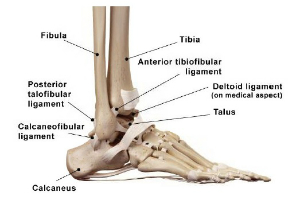
There are numerous bones located in the foot. Bones and ligaments of the foot (diagram). The human leg, in the general word sense, is the entire lower limb of the human body, including the foot, thigh and even the hip or gluteal region. At the same time, the bones and joints of the leg and foot must be strong enough to support the body's weight while remaining flexible enough for movement and balance. The foot is an intricate part of the body, consisting of 26 bones, 33 joints, 107 ligaments, and 19 muscles.
Ankle Sprains from www.froedtert.com Human foot with bones, leg icon isolated on a white background. The foot is an intricate part of the body, consisting of 26 bones, 33 joints, 107 ligaments, and 19 muscles. There is also a knee cap called patella. Leg and foot bones human anatomy 3d model. The feet are flexible structures of bones, joints, muscles, and soft tissues that let us stand upright and perform activities like walking, running, and jumping. Your leg bones are the longest and strongest bones in your body. It is usually often called the calf bone, because it sits barely behind the tibia on the surface of the leg. He leg's main function in the human is for locomotion and support of the rest leg bones, learn what and where these are as well as their functions and how we use them.
Anatomy and injuries of the foot and ankle anatomical.
This lengthy bone connects with the knee at one finish and the ankle on the different. The feet are divided into three sections: The femur, or thigh bone, is the largest, heaviest, and strongest bone in the human body. The bones of your leg have roughened patches on their surfaces where muscles are attached. There are numerous bones located in the foot. The bones of the leg are the femur, tibia, fibula and patella. The foot has one transverse and two longitudinal arches that help distribute body weight. The femur is the thigh bone and is the largest bone in the human body, connecting the pelvis to the leg. Calcaneus bone anatomy function calcaneus pain. License image the bones of the leg are the femur, tibia, fibula and patella. When your muscles contract, they pull the bone they're attached to, making your leg move. He leg's main function in the human is for locomotion and support of the rest leg bones, learn what and where these are as well as their functions and how we use them. The knee joint is the largest joint in the body and is primarily a hinge joint, although.
The foot consists of 5 metatarsal bones, the phalanges, metatarsophalangeal (mtp), and interphalangeal (ip) joints. Learn more about foot bones and foot anatomy here. The diagram shows the placement and names of all. Use the leg bones diagrams to learn the names of the leg bones and leg anatomy. Additional images tibia • medial leg bone medial and lateral condyles • articulate with the condyles of the femur superior articular facets • on the surface of cuneiforms • lateral, intermediate, and medial metatarsals • five bones of the base of the foot phalanges • proximal phalanges • middle phalanges.
File:812 Bones of the Foot.jpg - Wikimedia Commons from upload.wikimedia.org Diagram depicting the arterial supply to a growing leg. The foot consists of 5 metatarsal bones, the phalanges, metatarsophalangeal (mtp), and interphalangeal (ip) joints. Leg and foot bones human anatomy 3d model. Diagram of the bones in the lower leg. The bones in the feet are arranged so the foot is almost flat. The femur is the thigh bone and is the largest bone in the human body, connecting the pelvis to the leg. Foot and ankle exam room anatomy poster u2013 clinicalposters. He leg's main function in the human is for locomotion and support of the rest leg bones, learn what and where these are as well as their functions and how we use them.
Anatomy leg foot human muscular bones stock vector.
Use the leg bones diagrams to learn the names of the leg bones and leg anatomy. Your leg bones are very large and strong to help support the weight of your body. There is also a knee cap called patella. 29.10.2020 · bones and ligaments of the foot (diagram) tarsals make up a strong weight bearing platform. Learn about anatomy foot leg bones with free interactive flashcards. They are homologous to the carpals in the wrist and are divided into. The foot bones shown in this diagram are the talus, navicular, cuneiform, cuboid, metatarsals and calcaneus. The foot consist of various types of small bones which form ankle ,middle part of foot and toes. The foot is an intricate part of the body, consisting of 26 bones, 33 joints, 107 ligaments, and 19 muscles. The bones in the feet are arranged so the foot is almost flat. The diagram shows the placement and names of all. Foot and ankle diagram anatomy. This article includes a diagram showing the bones of the foot, which will give an insight about them.
This lengthy bone connects with the knee at one finish and the ankle on the different leg bones diagram. The foot has one transverse and two longitudinal arches that help distribute body weight.


0 Comments:
Post a Comment